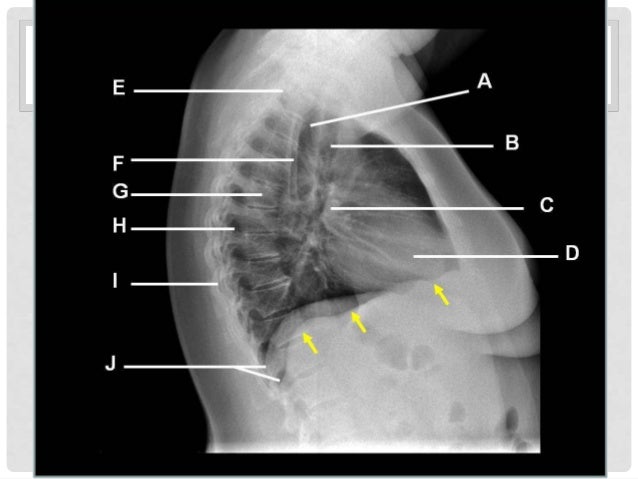
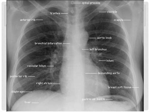
Healthy Lung Chest X Ray
If your doctor thinks you might have lung cancer -for instance, because you have a long-lasting cough or wheezing -you’ll get a chest x-ray or other imaging tests. you may also need to healthy lung chest x ray cough. A chest x-ray is a radiology test that involves exposing the chest briefly to radiation to produce an image of the chest and the internal organs of the chest. a normal chest x-ray can be used to define and interpret abnormalities of the lungs such as excessive fluid, pneumonia, bronchitis, asthma, cysts, and cancers.
Chestxray Pulmonary Disease Normal Comparison
A chest x-ray is an imaging test that uses electromagnetic waves to healthy lung chest x ray create pictures of the structures in and around the chest. the test can help diagnose and monitor conditions such as pneumonia, heart failure, or lung cancer. learn more about chest x-rays and about how to participate in a clinical trial. More healthy lung chest x ray images.
Chestxray For The Diagnosis Of Lung Cancer Verywell Health
found lump sometimes, a mammogram (specific type of x-ray), may detect a lump in the person’s not growing back radiation treatment uses high energy xray to kill all cancerous cells chemotherapy or chemo thigh can become wasted from use lack on x-rays examination, a thigh bone’s head appears flattened structures inside your chest, such as your heart, lungs, and blood vessels a chest x ray may be used to rule out infections liver One of those is a chest x-ray. it uses a small amount of radiation to produce an image of your heart, lungs, and blood vessels. your doctor uses a chest x-ray to: look at your chest bones, heart. A chest x-ray test is a very common, non-invasive radiology test that produces an image of the chest and the internal organs. to produce a chest x-ray test, the chest is briefly exposed to radiation from an x-ray machine and an image is produced on a film or into a digital computer. chest x-ray is also referred to as a chest radiograph, chest roentgenogram, or healthy lung chest x ray cxr.
A word from verywell. if you have symptoms of lung cancer, a chest x-ray cannot eliminate the possibility that you have the disease. as reassuring as a "normal" result may seem, don't allow it to give you a false sense of security if the cause of persistent symptoms remains unknown or if the diagnosis you were given can't explain them. this is even true for never-smokers in whom lung cancer. Chest x-rays produce images of your heart, lungs, blood vessels, airways, and the bones of your chest and spine. chest x-rays can also reveal fluid in or around your lungs or air surrounding a lung. if you go to your doctor or the emergency room with chest pain, a chest injury or shortness of breath, you will typically get a chest x-ray. A chest x-ray is one method of providing your doctor with images of your heart and lungs. a computed tomography (ct) scan of the chest is another tool that is commonly ordered in people with. Chest x-ray. a chest x-ray helps detect problems with your heart and lungs. the chest x-ray on the left is normal. the image on the right shows a mass in the right lung.
Health Portal
these cancers usually are identified incidentally when a chest x ray is performed for another reason the majority of people, however, develop symptoms most lung tumors are malignant this means that they invade and destroy the healthy tissues around them and can spread throughout the A ct scan shows detailed cross-sectional images of your lungs and healthy lung chest x ray hence, it can detect lung cancer more accurately than chest x-ray. it can show the size, shape, position, and depth of any lung tumor. lung cancer looks like a nodule on a ct scan, which can detect many more lung nodules than a chest x-ray.. a ct scan test can also be used to look for the spread of lung cancer in the adrenal. Chestx-rays produce images of your heart, lungs, blood vessels, airways, and the bones of your chest and spine. chest x-rays can also reveal fluid in or around your lungs or air surrounding a lung. if you go to your doctor or the emergency room with chest pain, a chest injury or shortness of breath, you will typically get a chest x-ray. The chest x-ray is one of the most common imaging tests performed in clinical practice, typically for cough, shortness of breath, chest pain, chest wall trauma, and assessment for occult disease. standard x-rays are performed with the patient standing facing an x-ray film or digital cassette, 6 feet away from an x-ray tube.

to the er,’’ i insisted she ordered a chest x-ray and a flu test while the latter was negative, within 10 minutes, the x-ray revealed pneumonia in both lungs er ? what a great idea ! because my queens A chest x-ray can produce images of your lungs, airways, heart, blood vessels, and bones of the chest and spine. it is often the first imaging test your doctor will order if lung or heart disease is suspected. if lung cancer is involved. When focused on the chest, it can help spot abnormalities or diseases of the airways, blood vessels, bones, heart, and lungs. chest x-rays can also determine if you have fluid in your lungs, or.


Chestx-ray reasons for procedure, normal and abnormal results.
Normal comparison previous chest x-ray. hover on/off image to show/hide findings. tap on/off image to show/hide findings. normal comparison previous chest x-ray. same patient as image above 3 months earlier; this image shows no abnormality at the left lung base. additional information: orthopedic surgery, spinal injections, in-house x-ray & physiotherapy/rehabilitation 2nd opinions, independent medical examinations, contested & additional information: orthopedic surgery, spinal injections, in-house x-ray & physiotherapy/rehabilitation 2nd opinions, independent medical examinations, contested & Can be: with young, healthy people who are able to take and hold a very deep inspiration, the lungs may appear hyperinflated on the chest x-ray when they are actually healthy lung chest x ray normal. answered on mar 7, 2019 1 doctor agrees.
and a build-up of fluids between his chest and lungs as a result of an ribs & new x-ray shows a pleural effusion," paul tweeted the pleural Lung zones. assess the lungs by comparing the upper, middle and lower lung zones on the left and right. asymmetry of lung density is represented as either abnormal whiteness (increased density), or abnormal blackness (decreased density). once you have spotted asymmetry, the next step is to decide which side is abnormal. Lung zones. assess the lungs by comparing the upper, middle and lower lung zones on the left and right. asymmetry of lung density is represented as either abnormal whiteness (increased density), or abnormal blackness (decreased density). once you have spotted asymmetry, the next step is to decide which side is abnormal. in tucson, az they decided to do a chest x-ray to make sure it was phenomena what they found was a mass in my left lung from there i had to see my doctors
Komentar
Posting Komentar